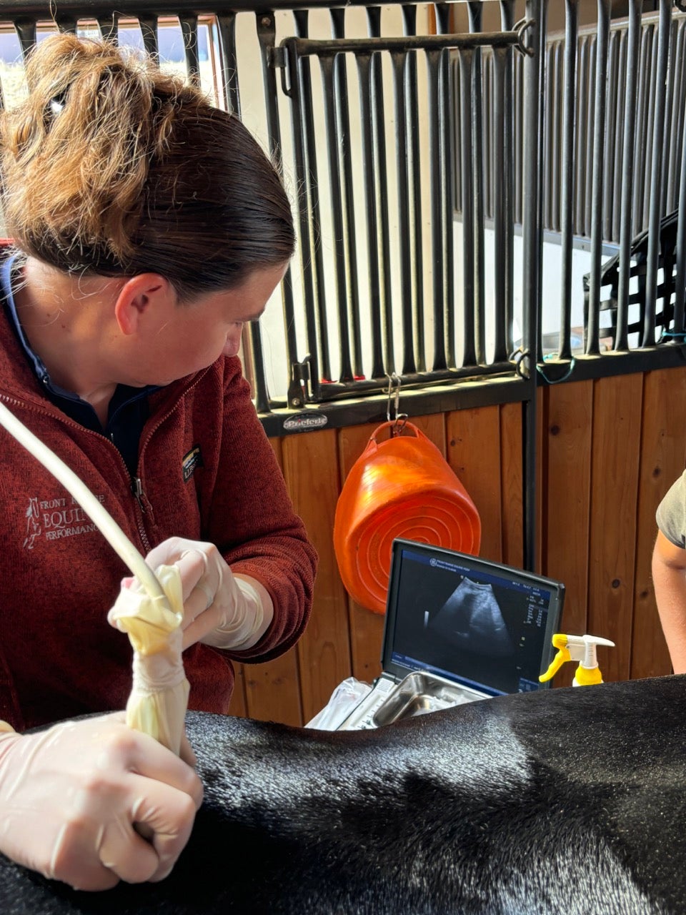
Ultrasonography

Diagnostic ultrasonography is a non-invasive, painless method used to identify and monitor soft tissue problems and to examine tendons, ligaments and muscles often related to lameness. It is often used in conjunction with other diagnostics to identify the root cause of a presenting concern.
In addition to diagnosis of the initial presenting concern, sequential (time-lapse) imaging is used to evaluate the progress of treatments over time, and is very useful in monitoring healing in musculoskeletal or soft tissue injuries so that controlled exercise can be accurately implemented. Ultrasonographic evaluation helps to characterize and document the size, shape, echogenicity (brightness) and fiber pattern of soft tissue. Ultrasound is also used to evaluate bony surfaces of regions that can not be otherwise imaged.
Front Range Equine’s portable ultrasound enables us to perform complete exams in the field and to archive or share the images with referring veterinarians and clients through wireless Internet.
Ultrasound is also often used in conjunction with critical injections or therapies, such as PRP, to more accurately deliver the therapy to the target area.
Ultrasound Guided Injections

Many ailments require specific placement of medications or regenerative therapies into a joint space, vessel, or directly into a tendon or ligament tear. Using an ultrasound, we are able to precisely guide medications to the specific area needing treatment.
Ultrasound guidance allows us to treat a variety of areas we cannot see or feel including:
-
Sacro-illiac joints
-
Cervical facets (neck joints)
-
Back joints (thoracic and lumbar facets)
-
Kissing spine
-
Soft tissue injuries (tendon, ligament, meniscus)
